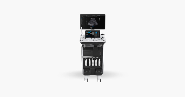
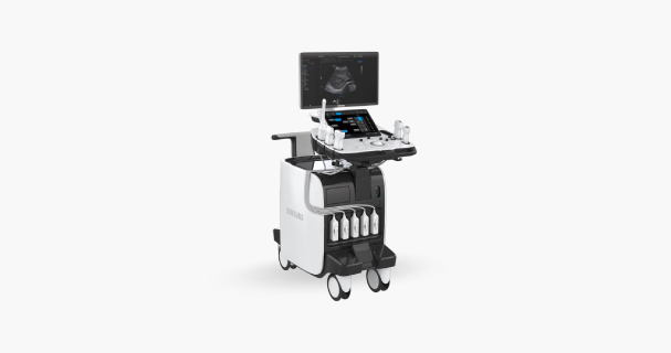
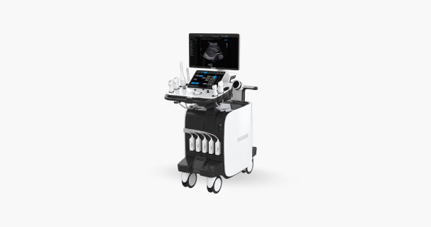
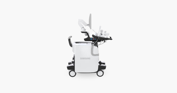
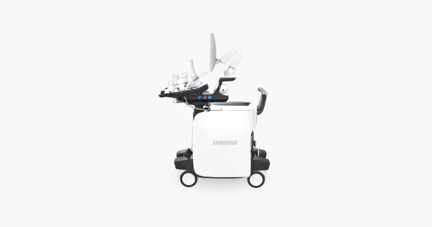
RS80A with Prestige
Volume rendering technology for accurate measurement
S-3D Arterial Analysis™ measures artery plaque volume and tracks morphological changes.
It helps accurate, early detection of cardiovascular diseases.
Evolving detection tool for lesion analysis
S-Detect™ for breast provides the characteristics of displayed lesion and a recommendation on whether the lesion is benign or malignant by adopting deep learning detection algorithm.
Features
Superior image quality
S-Vision Architecture
S-Vision Beamformer
The S-Vision Beamformer receives returning signals through a sophisticated digital filtering system resulting in reduced side lobes, less noise and artifact.
S-Vision imaging engine
The S-Vision Beamformer receives returning signals through a sophisticated digital filtering system resulting in reduced side lobes, less noise and artifact.
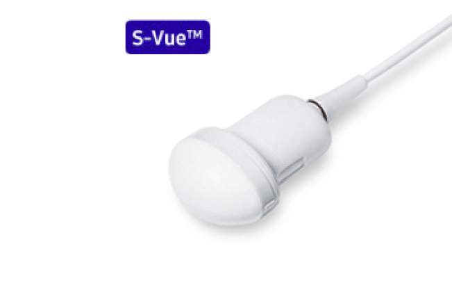
Imaging becomes Even Better with S-Vue transducer
(Single Crystal Technology)
The S-Vue transducers provide broader bandwidth and higher sensitivity over conventional Samsung transducers. They enable higher resolution at depth thereby providing improved image quality even with technically challenging patients.
In addition, the ergonomically designed S-Vue transducer fits well in the hand and is easy to handle.


S-Harmonic
This new harmonic technology provides greater image uniformity from near to far field while reducing signal noise.
HQ Vision
HQ Vision represents new, advanced technology for visualizing anatomical structures. It helps to make a reliable diagnosis quickly.
For advanced lesion diagnosis
CEUS+
CEUS+ technology uses the unique properties of ultrasound contrast agents. When stimulated with low MI frequencies, the oscillating microbubbles reflect both basic frequencies and harmonic signals.
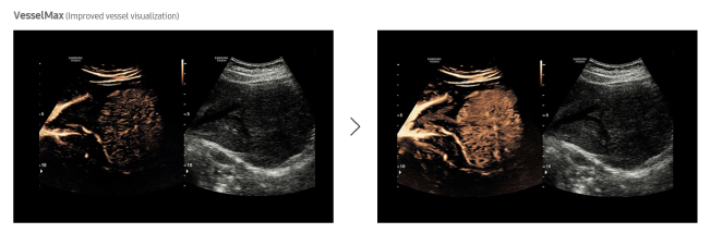
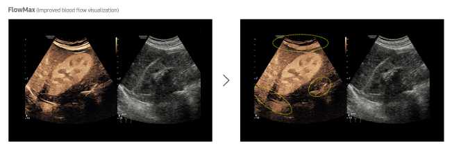
In addition, Samsung’s latest technologies, VesselMax and FlowMax, provide a clear visualization of vessels and blood flow so that you can form an informed, reliable diagnosis with confidence.

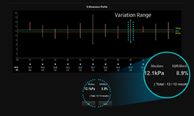
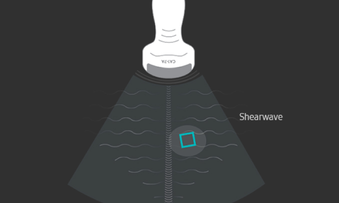
S-Shearwave
S-Shearwave detects the velocity of the shearwave propagated through the targeted lesion and displays the numerical measurement of stiffness In kPa or m/s together with a Reliable measurement Index (RMI)*. S-Shearwave has the potential to reduce the number of conventional liver biopsies by providing quantitative tissue characteristic information.
E-Strain
E-Strain enables quick and easy calculation of the strain ratio between two regions of interest in day-to-day procedures such as breast, prostate or gynecological examinations.
E-Breast™
Unlike conventional ultrasound elastography, E-Breast™requires to select only one ROI to calculate the strain ratio.
E-Thyroid™
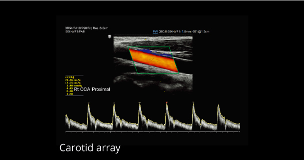
E-Thyroid™ uses pulsations from the adjacent Carotid Artery and provides an assessment of thyroid lesions.
S-Detect™ for Breast
S-Detect™ employs BI-RADS® scores for standardized analysis and classification of suspicious lesions. It provides the characteristics of displayed lesion and a recommendation on whether the lesion is benign or malignant. With 3 modes included in S-Detect™, users can set the level of sensitivity and specificity for a specific purpose.
S-Detect™ for Thyroid
S-Detect™ for Thyroid detects and classifies suspicious thyroid lesions semi-automatically based on Thyroid Image Reporting and Data System (TI-RADS) scores. This technology helps you diagnose your patients with confidence and ease, providing accurate, consistent results and an automatic reporting feature.
For advanced intervention procedure
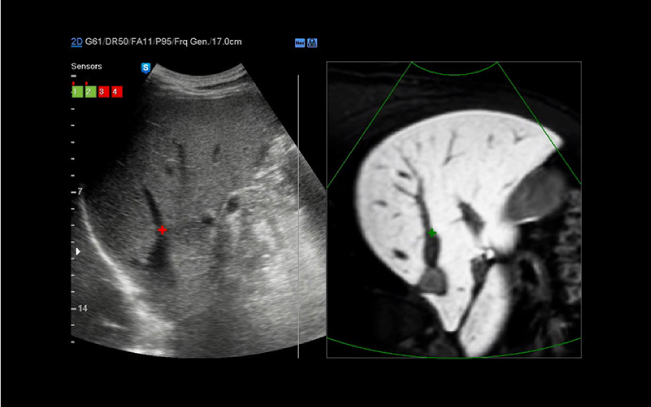
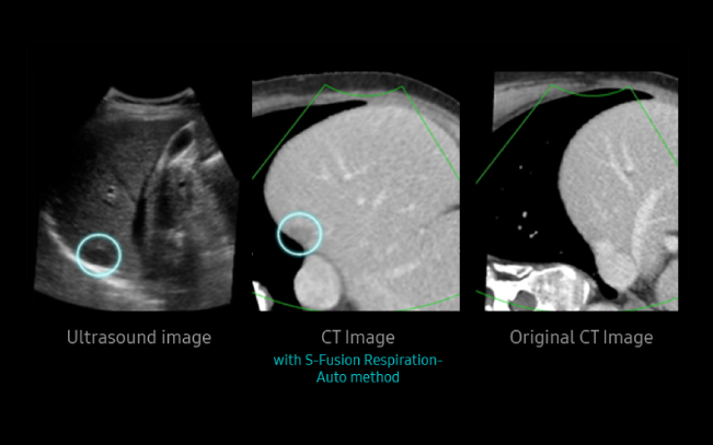
S-Fusion
S-Fusion enables simultaneous localization of a lesion with a real-time ultrasound in conjunction with other 3D volumetric imaging modalities. Unlike the conventional image fusion technology, Samsung offers a quicker and more precise registration process. S-Fusion enables precise targeting during operations such as Percutaneous Needle Biopsy and Radiofrequency Ablation.
Respiration Auto
S-Fusion’s Respiration Auto has been developed to minimize registration difference between the CT images and ultrasound scan images and provides more accurate anatomical images of liver without an influence of patient’s respiration. This technology can accurately generate virtual expiratory CT volume from patient’s inspiratory CT scan.
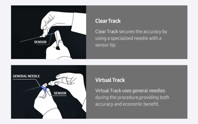
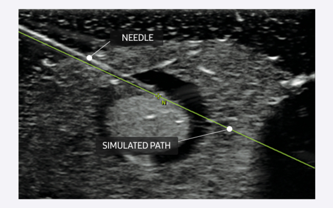
S-Tracking
S-Tracking increases the rate of accuracy during interventional procedures by providing the simulated path of the needle and the target mark in the live ultrasound image.
S-3D Arterial Analysis™
Based on the available 3D data, S-3D Arterial Analysis™ helps measure artery plaque volume for quantitative analysis purposes, as well as track morphological changes. This technology enables the accurate detection of cardiovascular diseases.
For cardiovascular disease
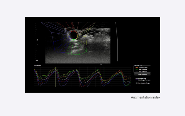
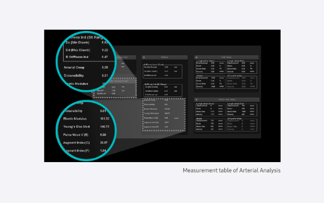
Arterial Analysis™
Arterial Analysis™ detects functional changes of vessels, providing measurement values such as the stiffness and intima-media thickness. Since the functional changes occur before morphological changes, this technology supports the early detection of cardiovascular diseases.
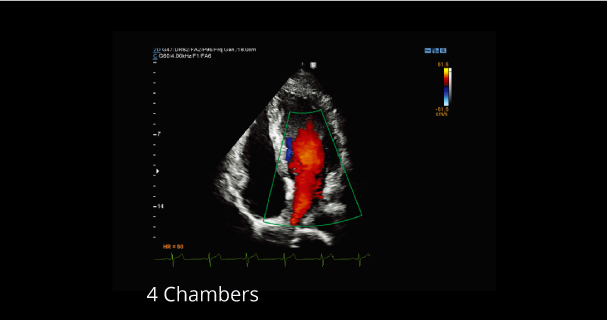
Strain+
Strain quantitatively displays a Bull’s Eye which shows left ventricular motion and dyssynchrony at a glance.
Stress Echo
The Stress Echo package includes wall motion scoring and reporting. It includes exercise Stress Echo, pharmacologic Stress Echo, diastolic Stress Echo and free programmable Stress Echo.
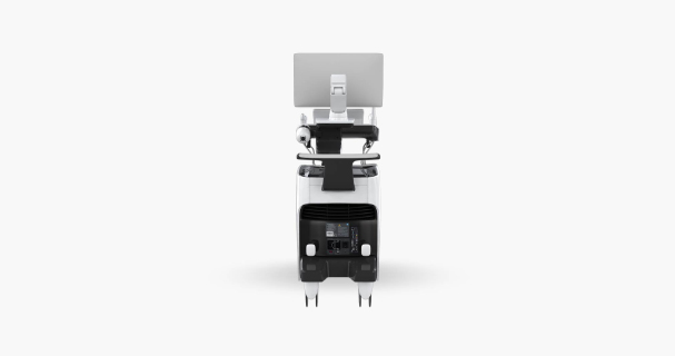
Design for your convenience
Folding monitor
The folding monitor enables safe and secure transport.

13.3-inch tilting touch screen
The tilting touch screen adjusts to accommodate user viewing preference in any scanning environment.
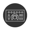
6 way adjustable control panel
The RS80A with Prestige’s 6 way adjustable control panel optimizes work environment to reduce repetitive stress. When off-mode, the control panel returns to home position for easier mobility.
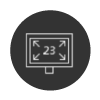
23-inch LED display
The RS80A with Prestige features a 23-inch high definition LED display delivering excellent contrast resolution, image clarity and vibrant color in any lighting condition.
Simplified console design
The simplified control panel including 3D Navigator and intuitive grouping of console buttons streamlines system interaction for efficient patient scanning.
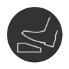
Swivel lock
A single pedal controls a swivel lock mechanism to conveniently secure console in place and accommodates efficient movement during a variety of scanning procedures.
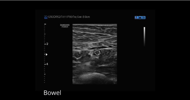
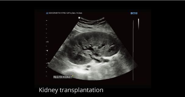
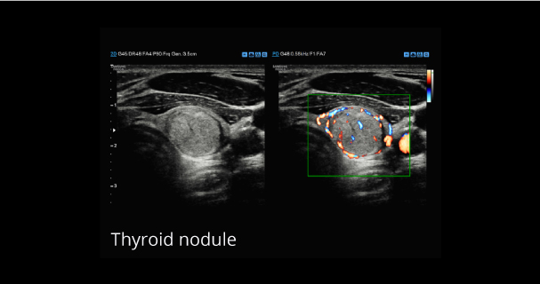
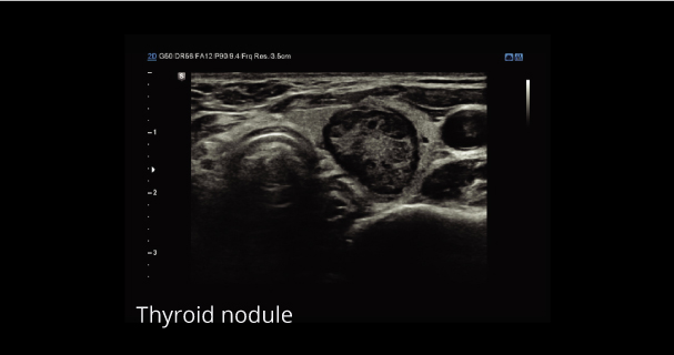
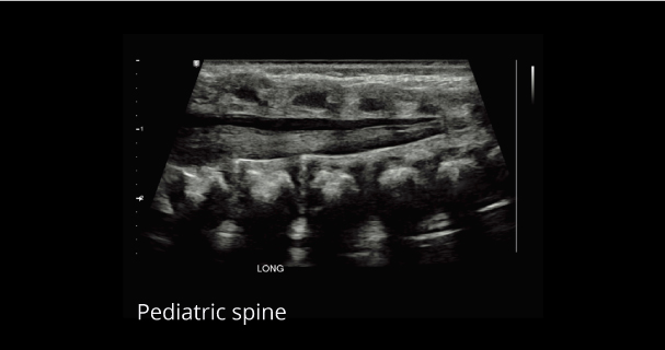
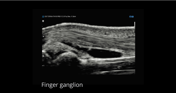
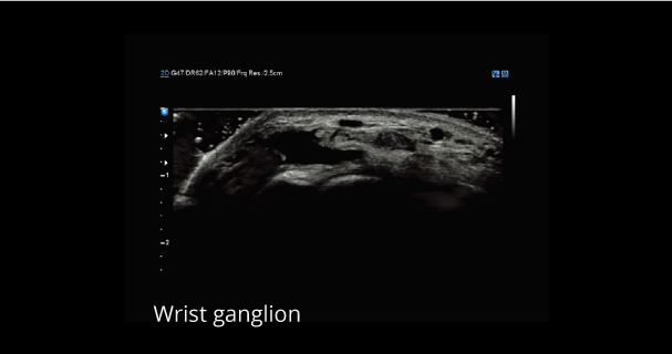
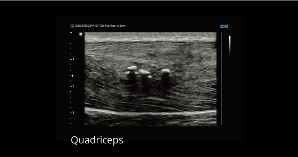
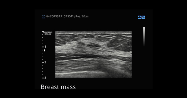
Clinical Images
Product Images
Videos
Documentation
| Information of Cleaning , Disnefectant and Ultrasound Gel | Download |
|---|
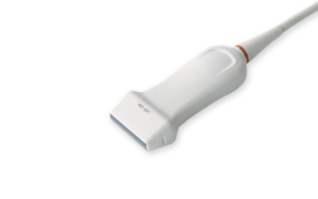
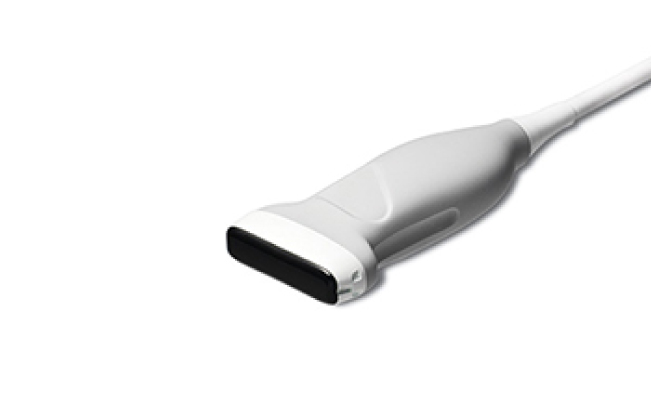
Transducers
Curved array transducers
CA1-7A
Application:
Abdomen, Obstetrics, Gynecology, Contrast
CA3-10A
Application:
Abdomen, Obstetrics, Gynecology, Pediatric, Vascular
CA2-8A
Application:
Abdomen, Obstetrics, Gynecology
CF4-9
Application:
Pediatric, Vascular
Linear array transducers
LM4-15B
Application:
Small parts, Vascular, Musculoskeletal
LA4-18B
Application:
Small parts, Vascular, Musculoskeletal
L3-12A
Application:
Small parts, Vascular, Musculoskeletal
LA3-16A
Application:
Small parts, Vascular, Musculoskeletal
LA2-9A
Application:
Small parts, Vascular, Musculoskeletal, Abdomen
LA3-16AI
Application:
Musculoskeletal
LA3-16A
Application:
Small parts, Vascular, Musculoskeletal
L7-16
Application:
Small parts, Vascular, Musculoskeletal
Volume transducers
LV3-14A
Application:
Musculoskeletal, Small parts, Vascular
V5-9
Application:
Obstetrics, Gynecology, Urology
CV1-8A
Application:
Abdomen, Obstetrics, Gynecology

V4-8
Application:
Abdomen, Obstetrics, Gynecology
Endocavity transducers
E3-12A
Application:
Obstetrics, Gynecology, Urology
Phased array transducers
PM1-6A
Application:
Cardiac, TCD, Abdomen
PA3-8B
Application:
Cardiac, Pediatric, Abdomen
PA4-12B
Application:
Cardiac, Pediatric
CW transducers
CW6.0
Application:
Cardiac
DP2B
Application:
Cardiac
TEE transducer
MMPT3-7
Application:
Cardiac